Where to find the BIC microscopes
On this page you will find your way to the microscopes via our floor plans as well as written directions.
Visiting us
The Biomedicum Imaging Core Facility (BIC) is located in the Biomedicum on the KI Campus in Solna. BIC’s microscopes can be found on the second floor (Rooms A0251/A0255) as well as the sixth floor (Rooms D0611/ D0613) of the Biomedicum building.
Visiting address: Solnavägen 9, 171 65 Solna
Facility email: bic@ki.se
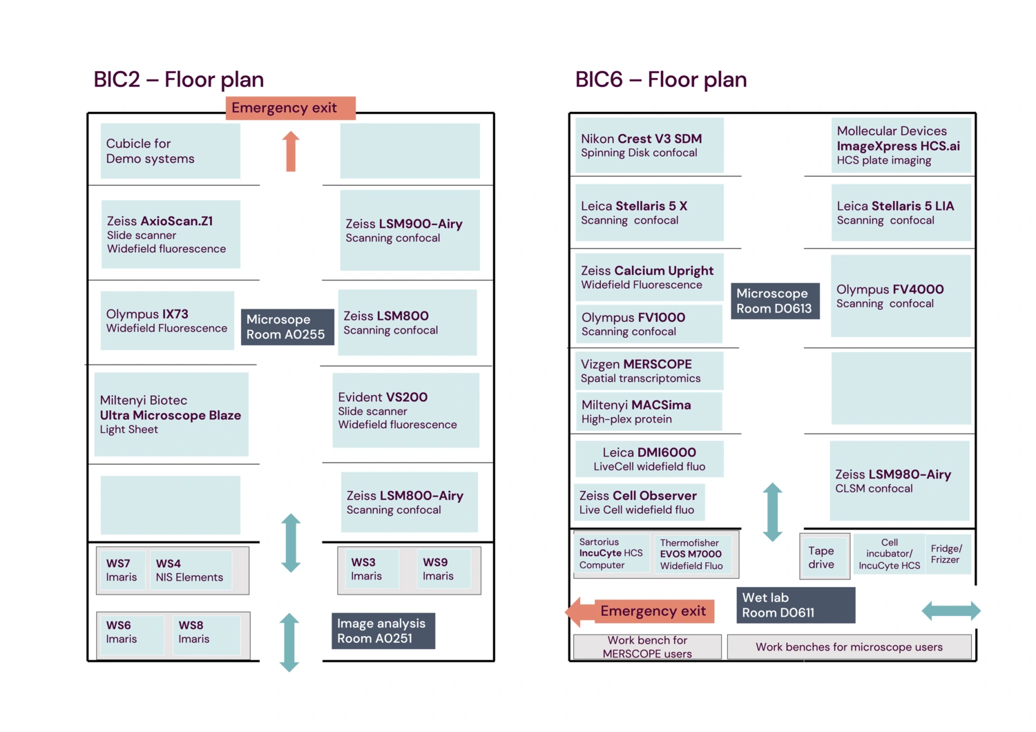
Download floor plans
BIC2
Image Analysis Room A0251
Entry to BIC2 is through Image Analysis Room A0251.
Equipment in A0251
Left hand side:
- WS8 (with Imaris and Amira)
- WS6 (with Imaris)
- WS7 (with Imaris)
- WS4 (with NIS Elements +ZEN)
Right hand side:
- WS3 (with Imaris)
- WS9 (with Imaris)
Microscope Room A0255
To reach the Microscope room A0255, enter via Image analysis room A0251, and proceed straight ahead.
Equipment in A0255
Left hand side (in the order from the entrance):
- Empty cubicle
- Miltenyi Ultra Microscope Blaze Light Sheet
- Olympus IX73 Widefield Fluorescence
- Zeiss AxioScan.Z1 Slide scanner
- Cubicle for Demo systems
Right hand side (in the order from the entrance):
- Zeiss LSM800-Airy Scanning confocal
- Evident VS200 Slide scanner
- Zeiss LSM800 Scanning confocal
- Zeiss LSM900-Airy Scanning confocal
- empty cubicle
Please note emergency exit is located at the back of the microscopy room A0255, opposite the entrance.
BIC6
Wet Lab Room D0611
Entry to BIC6 is through Wet Lab Room D0611.
Equipment in D0611
Left hand side:
- Work bench for the Wet Lab
- Work bench for MERSCOPE users (RNAse free)
Right hand side (in order from the entrance):
- Fridge/Frizzier
- Cell incubator / Sartirius IncuCyte S3 HCS
- Tape drive
- Entrance to the Microscope Room D0613
- ThermoFisher EVOS M700 Widefield Fluorescence
- Sartorius IncuCyte S3 HCS computer
Microscope room D0613
To reach the microscope room D0613, enter via Wet Lab Room D0611 and turn right to the Microscope room D0613
Equipment in D0613
Left hand side (in the order from the entrance):
- Zeiss Cell Observer Live Cell widefield fluorescence
- Leica DMI6000 Live Cell widefield fluorescence
- Vizgen MERSCOPE Spatial transcriptomics
- Miltenyi MACSima high-plex protein profiling
- Olympus FV1000 Scanning confocal
- Zeiss “Calcium upright” Widefield fluorescence
- Leica Stellaris 5 X Scanning confocal
- Nikon Crest SDM Spinning Disk confocal
Right hand side (in order from the entrance):
- Zeiss LSM980-Airy Scanning confocal
- Nikon Eclipse Ti2 Widefield fluorescent
- Olympus FV4000 Scanning confocal
- Leica Stellaris 5 LIA Scanning confocal
- Molecular Devices ImageExpress HCS.ai spinning disk confocal plate imaging
Please note that the emergency exit is located at the back of the Wet Lab Room D0611, opposite the entrance.